

💡 What You Need to Know Right Away
Doppler ultrasound is a medical imaging test that uses sound waves to measure blood flow through your arteries and veins, helping doctors detect blood clots, narrowed vessels, and circulation problems throughout your body.
Also known as: Duplex ultrasound, Vascular ultrasound, Doppler imaging, Color Doppler, Spectral Doppler
- Correctly identifies severe carotid artery narrowing in about 99 out of 100 cases[Evidence: A][1]
- Detects leg blood clots with 92-100% accuracy depending on the scanning method used[Evidence: A][7]
- Identifies portal vein blood clots in about 9 out of 10 cases[Evidence: A][4]
- Helps reduce baby deaths by about 29% when used to monitor high-risk pregnancies[Evidence: A][10]
If your doctor has recommended a Doppler ultrasound, you probably have questions. What exactly does this test measure? Will it hurt? How should you prepare? It's completely normal to feel uncertain about a medical test you haven't experienced before.
The good news is that Doppler ultrasound is one of the most patient-friendly diagnostic tests available. It uses no radiation, requires no needles, and typically takes less than an hour. Whether you're being checked for blood clots in your legs, narrowing in your neck arteries, or blood flow to your baby during pregnancy, this test provides critical information without discomfort.
In this guide, you'll learn exactly what happens during a Doppler ultrasound, how to prepare for your specific type of scan, what your results mean, and when this test is most useful. Our information comes from peer-reviewed research and clinical guidelines to give you accurate, trustworthy answers.
❓ Quick Answers
What is Doppler ultrasound used for?
Doppler ultrasound detects blood flow problems throughout your body. Doctors use it to find blood clots in legs (DVT), check for narrowed arteries in your neck that could cause stroke, monitor baby's blood flow during high-risk pregnancies, and evaluate circulation in your arms and legs[Evidence: A][8]. The test measures how fast blood moves and in which direction.
Is Doppler ultrasound safe?
Doppler ultrasound is safe and noninvasive with no radiation exposure. Unlike X-rays or CT scans, it uses only sound waves that bounce off moving blood cells. No health harms have been linked to diagnostic Doppler when used correctly. The test is safe during pregnancy when exposure time is kept reasonable[Evidence: A][10].
How does Doppler ultrasound work?
Doppler ultrasound works by sending sound waves into your body that bounce off moving blood cells. When blood flows toward the device, the returning sound waves have a higher frequency. When blood flows away, the frequency is lower. This "Doppler effect" lets doctors measure blood speed and direction[Evidence: B][5].
Is Doppler ultrasound painful?
Doppler ultrasound is not painful. You may feel light pressure as the sonographer moves the handheld device (transducer) across your skin. The water-based gel applied to your skin may feel cold initially but warms quickly. Some people hear a whooshing sound from the machine, which is normal and represents your blood flow.
How long does a Doppler ultrasound take?
A standard Doppler ultrasound takes 30 to 60 minutes depending on which body area is examined. Simple single-vessel checks may take only 15 to 20 minutes. Comprehensive vascular studies or echocardiograms with Doppler can take 60 to 90 minutes. Your appointment letter should specify the expected duration.
What is the difference between Doppler ultrasound and regular ultrasound?
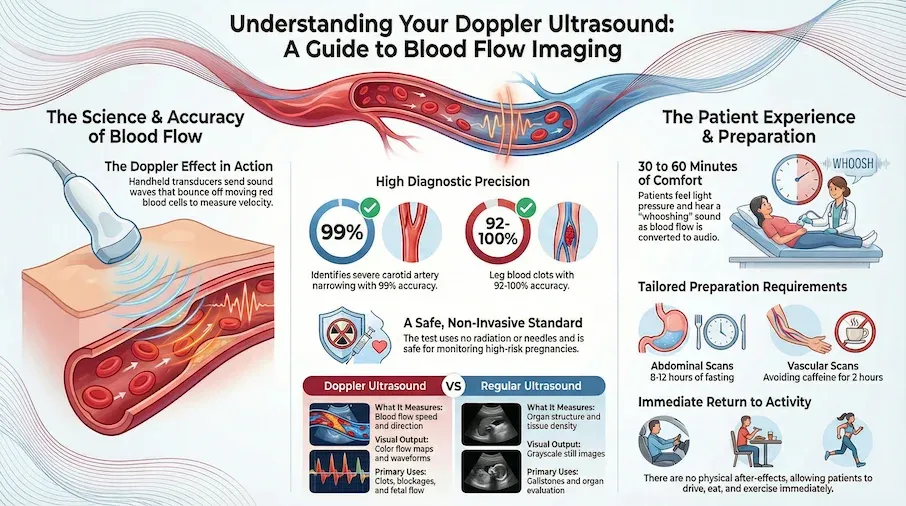
Regular ultrasound creates still images of organs and tissues, while Doppler ultrasound specifically measures blood movement. Regular ultrasound shows what structures look like. Doppler shows how blood flows through them, including speed, direction, and whether any blockages exist. Many scans combine both techniques (duplex ultrasound).
Do I need to prepare for a Doppler ultrasound?
Preparation depends on which body area is being scanned. Abdominal Doppler may require fasting for 8-12 hours. Vascular scans of arms or legs typically need no fasting but may require avoiding caffeine for 2 hours. Wear loose, comfortable clothing. Your clinic will provide specific instructions based on your scan type.
Doppler Ultrasound
A non-invasive medical imaging technique used to estimate the blood flow through your blood vessels by bouncing high-frequency sound waves off circulating red blood cells.
🔬 How Does Doppler Ultrasound Work?
Doppler ultrasound works like an echo bouncing off moving objects. Imagine standing near a highway and listening to a car horn. As the car approaches, the horn sounds higher-pitched. As it drives away, the pitch drops. This change in sound frequency based on motion is called the Doppler effect, named after physicist Christian Doppler who described it in 1842.
During your scan, a handheld device called a transducer sends high-frequency sound waves into your body. These sound waves bounce off your red blood cells as they travel through arteries and veins. When blood cells move toward the transducer, the returning sound waves have a slightly higher frequency. When they move away, the frequency is lower. The ultrasound machine measures these tiny frequency shifts and converts them into images and sounds.
Think of the transducer as a traffic camera that can see how fast cars are moving and which direction they're going. Instead of photographing license plates, it's measuring blood cell velocity. Research shows this technique correctly identifies severe carotid artery narrowing in about 99 out of 100 cases when the narrowing is present[Evidence: A][1].
Different types of Doppler provide different information:
- Color Doppler overlays colors on the image to show blood flow direction (typically red for flow toward the transducer, blue for flow away)
- Spectral Doppler displays blood velocity as a waveform graph, showing flow patterns over time
- Power Doppler is more sensitive to slow blood flow and helps visualize tiny blood vessels
- Duplex ultrasound combines regular ultrasound images with Doppler flow information
For detecting deep vein blood clots, research shows duplex ultrasound achieves 100% sensitivity and 91.7% specificity in certain settings[Evidence: B][5]. A large meta-analysis of 43 studies found proximal compression ultrasound has 90.1% sensitivity and 98.5% specificity for DVT in the upper leg[Evidence: A][8].
🧪 What to Expect: The Real User Experience
During the Procedure
When you arrive for your Doppler ultrasound, you'll lie on an examination table, typically on your back. The sonographer will apply a water-based gel to your skin over the area being examined. This gel feels cold when first applied (around 65-70°F) but warms to body temperature within 30-60 seconds. The gel helps sound waves travel between the transducer and your body.
As the sonographer moves the transducer across your skin, you'll feel light to moderate pressure. For deeper vessels like the abdominal aorta or in patients with higher body mass, the pressure may be slightly firmer, but this is typically described as "pressing" rather than painful. You may need to turn from side to side during limb scans, and for carotid scans, you'll turn your head to the side, which may cause mild neck tension if you have stiffness.
The distinctive "whooshing" sound is what many patients remember most. This rhythmic, pulsing sound is your actual blood flow being converted to audio. The pitch changes indicate blood flow speed and direction. Higher pitches mean faster flow toward the transducer. Lower pitches mean flow away. Many patients find listening to their own blood flow educational and even reassuring.
What You'll Feel After
There are no physical after-effects from a Doppler ultrasound. You won't experience soreness, bruising, or fatigue. The gel wipes off easily with a towel, though some residue may remain on your skin or clothing. You can resume all normal activities immediately, and there are no dietary restrictions afterward. The most common "effect" reported is simply anxious waiting for results.
How to Make It Easier
- For the cold gel: Ask the sonographer if they can warm the gel packet in their hands before applying
- For easy access: Wear two-piece clothing so you can expose the scan area without fully undressing
- If the sounds cause anxiety: Bring headphones, though most patients find the whooshing sounds reassuring
- For carotid scans: If you have neck stiffness, practice turning your head to the side beforehand
- To stay engaged: Ask to see the screen during scanning. Many patients find watching educational and calming
- For pressure discomfort: Communicate with your sonographer immediately. They can adjust their technique
📊 Procedure Details and How to Prepare
Your preparation requirements depend on which area of your body is being scanned. Use this table to understand what's expected:
| Scan Type | Fasting Required | Clothing | Other Restrictions | Duration |
|---|---|---|---|---|
| Abdominal (aorta, renal, mesenteric) | Yes, 8-12 hours before | Two-piece; loose waistband | No gum or smoking day of test | 30-45 min |
| Carotid (neck arteries) | No | No necklaces; open collar | Avoid caffeine 2 hours before | 30-45 min |
| Lower extremity (leg veins/arteries) | No | Shorts or loose pants | Avoid caffeine 2 hours before | 30-60 min |
| Upper extremity (arm veins/arteries) | No | Short sleeves or removable top | Avoid caffeine 2 hours before | 30-45 min |
| Obstetric (fetal/umbilical) | No (full bladder sometimes) | Two-piece; easy abdominal access | None typically | 30-45 min |
| Echocardiogram with Doppler | No | Hospital gown usually provided | None typically | 60-90 min |
General Preparation Guidelines
- Bring your medication list: Especially note blood thinners (warfarin, apixaban, rivaroxaban) as these affect interpretation
- Arrive 15 minutes early: Allow time for paperwork and changing if needed
- Leave jewelry at home: Necklaces interfere with carotid scans; rings and bracelets may need removal for arm scans
- Bring your referral: Some facilities require the original order from your doctor
- Ask about results timeline: Most facilities have results within 24-48 hours sent to your ordering physician
⚠️ Safety Information
The test itself is painless and involves no radiation exposure. Unlike CT scans or X-rays, Doppler ultrasound uses only sound waves, making it one of the safest imaging modalities available. Most people tolerate the procedure well, and there are no documented health harms from diagnostic Doppler when used according to standard protocols.
It's normal to feel anxious about any medical test, especially when you're waiting for results that could reveal a health problem. The anxiety about results is often more challenging than the test itself.
Special Considerations for Pregnancy
Research shows using Doppler ultrasound in high-risk pregnancies helped reduce baby deaths by about 29 percent[Evidence: A][10]. The same research found it helped reduce cesarean deliveries by about 10 percent. However, routine spectral Doppler in the first trimester is generally avoided due to higher acoustic output. Your obstetrician will determine the appropriate timing and type based on your specific situation.
After Your Test
You can drive yourself home, return to work, eat normally, and exercise without restrictions. If you have concerning symptoms that prompted the test (leg swelling, sudden vision changes, chest pain), continue following your doctor's guidance while awaiting results. If symptoms worsen, seek immediate medical attention regardless of pending ultrasound results.
🥗 Practical Day-of-Test Tips
How to Use This Information
Morning Appointment (Abdominal Scan)
- Night before: Eat a light dinner, finish eating by 8 PM
- Morning of: No food, no gum, no smoking; water only is typically allowed
- Clothing: Loose pants with elastic waistband, two-piece outfit
- Timing: Arrive 15 minutes early; expect 30-45 minutes for the scan
- After: Eat immediately after if hungry; bring a snack
Afternoon Appointment (Leg or Arm Scan)
- Morning: Eat normally; avoid caffeine 2 hours before appointment
- Clothing: Shorts or loose pants for leg scans; short sleeves for arm scans
- Timing: Arrive 15 minutes early; expect 30-60 minutes for the scan
- After: Resume all normal activities immediately
Common Mistakes to Avoid
- Wearing one-piece clothing: You'll need to expose the scan area, which is awkward in dresses or jumpsuits
- Drinking coffee before vascular scans: Caffeine affects blood vessel dilation and can alter results
- Forgetting your medication list: Blood thinners especially affect result interpretation
- Applying lotion to the scan area: Lotion can interfere with gel conductivity; skip it day-of
- Rushing to the appointment: Stress and hurrying elevate blood pressure, potentially affecting readings
What to Look for When Choosing a Doppler Ultrasound Provider
The quality of your test results depends significantly on the lab and healthcare provider. Here's what to consider:
Lab Quality Markers
- CLIA certification: Clinical Laboratory Improvement Amendments accreditation required for all diagnostic labs Why it matters: Federal quality standards ensure accurate results
- ICAVL accreditation: Intersocietal Accreditation Commission for vascular labs (gold standard for vascular ultrasound) Why it matters: Demonstrates rigorous quality protocols specific to vascular testing
- Sonographer credentials: Look for RDMS (Registered Diagnostic Medical Sonographer) or RVT (Registered Vascular Technologist) Why it matters: Certified technologists have verified training and ongoing competency
- Turnaround time: Most facilities provide results within 24-48 hours Why it matters: Faster results enable quicker treatment decisions, especially for suspected blood clots
- Insurance acceptance: Verify in-network status before scheduling Why it matters: Out-of-network facilities can cost 3-5 times more
Questions to Ask Your Provider
- Is your vascular lab ICAVL-accredited?
- What credentials do your sonographers hold?
- How long will results take?
- Will the interpreting physician be a board-certified radiologist or vascular specialist?
- What specific preparation do I need for my type of scan?
- What's my out-of-pocket cost if my insurance doesn't cover this?
Red Flags to Avoid
- No accreditation information available: Reputable facilities display credentials
- Unusually cheap pricing: May indicate outdated equipment or unlicensed staff
- Results take more than a week: Standard turnaround is 24-48 hours
- No radiologist interpretation: Images should be read by a licensed physician
How Doppler Ultrasound Compares to Regular Ultrasound: What to Know
Doppler ultrasound and regular ultrasound use the same basic technology (sound waves) but provide different types of information. Regular ultrasound creates still images of organs and structures. Doppler adds the ability to measure blood flow. Many modern scans combine both techniques in what's called duplex ultrasound.
| Feature | Doppler Ultrasound | Regular Ultrasound |
|---|---|---|
| What It Measures | Blood flow speed, direction, and vessel patency | Organ structure, tissue density, masses |
| Image Type | Color flow maps + waveform graphs + audio | Grayscale still images or real-time video |
| Primary Uses | Blood clot detection, artery narrowing, fetal blood flow monitoring | Pregnancy imaging, gallbladder/kidney stones, organ evaluation |
| Sound During Exam | Yes (whooshing blood flow sounds) | Usually silent or minimal |
| Typical Duration | 30-60 minutes | 15-30 minutes |
| Radiation Exposure | None (sound waves only) | None (sound waves only) |
When Each Type Is Appropriate
Choose Doppler when: Your doctor suspects blood flow problems. Blood clots in legs (DVT), stroke prevention screening (carotid arteries), monitoring high-risk pregnancy, checking circulation after vascular surgery.
Choose regular ultrasound when: Your doctor needs to see organ structure. Pregnancy dating, gallstones, kidney problems, thyroid nodules, breast lumps.
Duplex (combined) is common for: Most vascular evaluations combine both techniques automatically. Your provider orders the appropriate type based on your clinical situation.
What The Evidence Shows (And Doesn't Show)
What Research Suggests
- Strong evidence (6 meta-analyses) supports high diagnostic accuracy for blood clot detection, with 90-100% sensitivity depending on scanning method[Evidence: A][7][8]
- For carotid stenosis grading, peak systolic velocity ≥230 cm/sec indicates ≥70% narrowing with 99% sensitivity across multiple validated centers[Evidence: A][1]
- In high-risk pregnancies, umbilical artery Doppler reduces perinatal deaths by 29% (NNT 203) and cesarean sections by 10%[Evidence: A][10]
- Portal vein thrombosis detection shows 89-93% sensitivity and 92-99% specificity[Evidence: A][4]
- When negative whole-leg ultrasound is obtained, only 1% of patients develop subsequent DVT[Evidence: A][9]
What's NOT Yet Proven
- Universal third-trimester umbilical artery screening shows only 21.7% sensitivity for detecting small babies[Evidence: A][11]. Benefits are clearer in HIGH-RISK pregnancies, not routine screening
- Calf vein (below-knee) DVT detection is less reliable than proximal DVT detection; some small clots may be missed
- Accuracy data for peripheral arterial disease and renal artery stenosis were not included in this evidence review
- Long-term outcomes beyond the immediate diagnostic period are less well-characterized for most applications
Where Caution Is Needed
- Operator dependence: Accuracy varies with sonographer training and experience. ICAVL-accredited labs maintain quality standards
- Body habitus: Imaging is more challenging in patients with higher body mass; deeper vessels may be harder to visualize clearly
- Recent surgery or trauma: Swelling and post-operative changes can affect interpretation
- First trimester pregnancy: Spectral Doppler avoided due to theoretical thermal concerns; timing determined by obstetrician
Should YOU Get This Test?
Best suited for: People with suspected blood clots (leg swelling, pain), stroke risk assessment (carotid evaluation), high-risk pregnancy monitoring, and post-vascular surgery follow-up. Most useful when clinical suspicion is moderate to high.
Not recommended for: Universal screening in healthy populations without risk factors. Evidence does not support routine use in low-risk pregnancy for detecting growth restriction.
Realistic timeline: Results typically available within 24-48 hours. Treatment decisions (e.g., blood thinners for DVT) often begin immediately based on ultrasound findings.
When to consult a professional: If you have leg swelling, leg pain, shortness of breath, transient vision changes, or concerns about fetal movement. Your doctor determines whether Doppler ultrasound is the appropriate test for your specific symptoms.
Frequently Asked Questions
How much does a Doppler ultrasound cost?
Costs vary significantly by location, facility type, and insurance coverage. With insurance, out-of-pocket costs typically range from $50-$300 depending on your plan's copay and deductible status. Without insurance, a standard venous Doppler can range from $200-$500 at outpatient imaging centers, while hospital-based scans may cost $1,000-$3,000. Echocardiograms with Doppler are generally more expensive, ranging $1,500-$3,800 without insurance. Medicare covers medically necessary ultrasounds with typical 20% coinsurance after deductible. Always verify costs with your specific facility before scheduling.
What can a Doppler ultrasound detect?
Doppler ultrasound detects conditions affecting blood flow. For deep vein blood clots (DVT), research shows it correctly identifies clots in 90-100% of cases depending on the scanning method. For carotid artery narrowing that could lead to stroke, the test identifies severe narrowing with 99% sensitivity. It also detects portal vein blood clots in about 9 out of 10 cases, aneurysms, arterial blockages, and abnormal blood flow in pregnancy.
What are the types of Doppler ultrasound?
Four main types exist: Color Doppler shows blood flow direction using colors (red toward transducer, blue away). Spectral Doppler displays velocity as a waveform graph and provides precise speed measurements. Power Doppler is more sensitive to slow blood flow and helps visualize tiny vessels. Duplex ultrasound combines regular grayscale imaging with Doppler flow information. Most modern vascular labs use duplex scanning as standard. Continuous wave Doppler and pulsed wave Doppler are variations used for different clinical situations.
Can Doppler ultrasound detect blood clots?
Yes, detecting blood clots is one of its primary uses. Research involving 26 studies found the simplified 2-point version correctly identified leg blood clots in about 92 out of 100 cases, with 97% accuracy for ruling them out. Complete compression ultrasound achieved 100% sensitivity. When a whole-leg scan was negative, blood clots were later found in only about 1 out of 100 people. For proximal DVT specifically, early research suggests 96% sensitivity and 96% specificity.
Is Doppler ultrasound safe during pregnancy?
Yes, when used appropriately. Research shows using Doppler to monitor high-risk pregnancies helped reduce baby deaths by about 29 percent and cesarean deliveries by about 10 percent. One baby death was prevented for every 203 women monitored. Studies suggest checking blood flow every two weeks works as well as checking weekly for preventing intensive care admissions. Spectral Doppler is generally avoided in the first trimester due to theoretical thermal concerns; your obstetrician determines appropriate timing.
What is a carotid Doppler ultrasound?
A carotid Doppler specifically examines the carotid arteries in your neck, which supply blood to your brain. Doctors order this test to check for atherosclerosis (plaque buildup) that could cause stroke. Research shows this test correctly identified severe carotid artery narrowing (70% or greater) with 99% sensitivity and 86% specificity. The sonographer measures blood flow velocity; faster speeds indicate narrower vessels. No fasting is required, though you should avoid caffeine 2 hours before the test.
What happens if Doppler ultrasound results are abnormal?
Abnormal results trigger follow-up actions depending on what was found. For blood clots, treatment typically begins immediately (blood thinners). For carotid narrowing, your doctor may recommend lifestyle changes, medications, or referral to a vascular surgeon depending on severity. For fetal concerns, closer monitoring or early delivery may be planned. Studies show that Doppler findings help guide early delivery decisions for small babies at risk. Your doctor will explain your specific results and next steps.
Can I eat before a Doppler ultrasound?
It depends on which area is being scanned. For abdominal Doppler (aorta, mesenteric, renal arteries), fasting for 8-12 hours is typically required because food causes gas and bowel activity that obscures images. For all other Doppler scans (carotid, leg, arm, fetal), you can eat normally. However, you should avoid caffeine 2 hours before vascular scans as it affects blood vessel dilation. Your scheduling instructions should specify fasting requirements; call the facility if unclear.
What should I wear to a Doppler ultrasound?
Wear loose, comfortable, two-piece clothing that allows easy access to the scan area. For carotid scans, wear an open-collar shirt and remove necklaces. For leg scans, wear shorts or loose pants that can be rolled up to the thigh. For arm scans, wear short sleeves. For abdominal scans, wear pants with an elastic waistband. Avoid jewelry near the scan area. Some facilities provide gowns, but arriving in appropriate clothing makes the process faster and more comfortable.
What is the difference between Doppler and duplex ultrasound?
Duplex ultrasound combines regular grayscale imaging with Doppler flow measurement in a single examination. 'Doppler' refers specifically to the blood flow measurement component. 'Duplex' means you're getting both the structural image AND the flow information together. In practice, most vascular labs perform duplex scanning as standard, and the terms are often used interchangeably. When your doctor orders a 'Doppler' of your legs, they typically receive a duplex study showing both vessel structure and blood flow characteristics.
Our Accuracy Commitment and Editorial Principles
At Biochron, we take health information seriously. Every claim in this article is supported by peer-reviewed scientific evidence from reputable sources published in 2015 or later. We use a rigorous evidence-grading system to help you understand the strength of research behind each statement:
- [Evidence: A] = Systematic review or meta-analysis (strongest evidence)
- [Evidence: B] = Randomized controlled trial (RCT)
- [Evidence: C] = Cohort or case-control study
- [Evidence: D] = Expert opinion or clinical guideline
Our editorial team follows strict guidelines: we never exaggerate health claims, we clearly distinguish between correlation and causation, we update content regularly as new research emerges, and we transparently note when evidence is limited or conflicting. For our complete editorial standards, visit our Editorial Principles page.
This article is for informational purposes only and does not constitute medical advice. Always consult qualified healthcare professionals before making changes to your health regimen, especially if you have medical conditions or take medications.


References
- 1 . Accuracy of the Society of Radiologists in Ultrasound (SRU) Carotid Doppler Velocity Criteria for Grading North American Symptomatic Carotid Endarterectomy Trial (NASCET) Stenosis: A Meta-Analysis, Journal of Ultrasound in Medicine, 2023;42(7):1423-1435. PubMed [Evidence: A]
- 2 . Doppler ultrasound of umbilical and middle cerebral artery in third trimester small-for-gestational age fetuses to decide on timing of delivery for suspected fetal growth restriction: A cohort with nested RCT (DRIGITAT), BJOG, 2024;131(8):1042-1053. PubMed [Evidence: B]
- 3 . Frequency of third trimester umbilical artery Doppler for improving neonatal outcomes in high-risk pregnancies: a randomized trial, Obstetrics & Gynecology Science, 2023;66(3):161-168. PubMed [Evidence: B]
- 4 . Diagnostic accuracy of Doppler ultrasound, CT and MRI in Budd Chiari syndrome: systematic review and meta-analysis, British Journal of Radiology, 2020;93(1109):20190847. PubMed [Evidence: A]
- 5 . Doppler ultrasound imaging for detection of deep vein thrombosis in plastic surgery outpatients: a prospective controlled study, Aesthetic Surgery Journal, 2015;35(2):204-14. PubMed [Evidence: B]
- 6 . Clinical Presentation, Management, and Outcomes of Deep Vein Thrombosis Based on Doppler Ultrasonography Examination, Angiology, 2016;67(6):587-95. PubMed [Evidence: C]
- 7 . Deep venous thrombosis (DVT) diagnostics: gleaning insights from point-of-care ultrasound (PoCUS) techniques in emergencies: a systematic review and meta-analysis, The Ultrasound Journal, 2024;16(1):37. PubMed [Evidence: A]
- 8 . Diagnosis of deep vein thrombosis of the lower extremity: a systematic review and meta-analysis of test accuracy, Blood Advances, 2020;4(7):1250-1264. PubMed [Evidence: A]
- 9 . Diagnostic accuracy of three ultrasonography strategies for deep vein thrombosis of the lower extremity: A systematic review and meta-analysis, PLoS ONE, 2020;15(2):e0228788. PubMed [Evidence: A]
- 10 . Fetal and umbilical Doppler ultrasound in high-risk pregnancies, Cochrane Database of Systematic Reviews, 2017;6:CD007529. PubMed [Evidence: A]
- 11 . Fetal umbilical artery Doppler as a tool for universal third trimester screening: A systematic review and meta-analysis of diagnostic test accuracy, Placenta, 2021;108:47-54. PubMed [Evidence: A]
Medical Disclaimer
This content is for informational and educational purposes only. It is not intended to provide medical advice or to take the place of such advice or treatment from a personal physician. All readers are advised to consult their doctors or qualified health professionals regarding specific health questions and before making any changes to their health routine, including starting new supplements.
Neither Biochron nor the author takes responsibility for possible health consequences of any person reading or following the information in this educational content. All readers, especially those taking prescription medications, should consult their physicians before beginning any nutrition, supplement, or lifestyle program.
If you have a medical emergency, call your doctor or emergency services immediately.
