

💡 What You Need to Know Right Away
CT urography is an imaging test that uses X-rays and contrast dye to create detailed pictures of your kidneys, ureters, and bladder, helping doctors detect kidney stones, tumors, infections, and causes of blood in urine.
Also known as: CT urogram, CTU, Computerized tomography urogram, CT IVP, Multidetector CT urography
- Detects kidney stones with about 93% accuracy and correctly rules them out in about 97% of cases[Evidence: A][11]
- Correctly identifies kidney cancer and upper urinary tract cancer in about 94 out of 100 cases when cancer is present[Evidence: A][1]
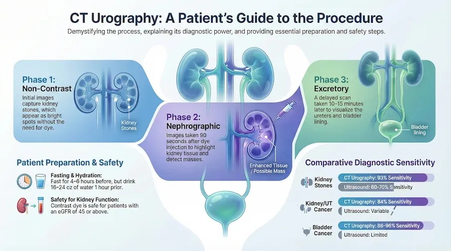
- The contrast dye does not increase kidney injury risk for people with mildly reduced kidney function (eGFR 45 or above)[Evidence: A][13]
- The entire appointment takes 45-60 minutes, but actual scan time is only 3-5 minutes
If your doctor has recommended a CT urography, you may be wondering what the test involves and what it can tell you about your health. It's common to feel anxious about medical imaging tests, especially if you've heard concerns about contrast dye or radiation exposure.
The good news is that CT urography is one of the most reliable tests for evaluating problems in your urinary tract. Whether you're being tested because of blood in your urine, kidney stones, or another concern, this guide will help you understand exactly what to expect before, during, and after your scan. We'll cover the science behind the test, how to prepare, what the experience feels like, and how to understand your results.
❓ Quick Answers
What is CT urography used for?
CT urography is used to evaluate blood in the urine (hematuria), detect kidney stones, find tumors in the kidneys or bladder, and diagnose infections or blockages in the urinary tract. Clinical guidelines recommend it as the primary imaging test for people being evaluated for blood in their urine[Evidence: D][4].
Is CT urography safe?
CT urography is safe for most people. Research shows that for people with mildly reduced kidney function (eGFR 45 or above), there is no increased risk of kidney injury from the contrast dye[Evidence: A][13]. The radiation exposure is minimized with modern techniques. Your doctor will check your kidney function before the test.
How long does a CT urography take?
The entire CT urography appointment takes 45-60 minutes, including check-in, IV placement, and the scan itself. The actual scanning time is only 3-5 minutes. You'll lie still on a table while the scanner takes images in three phases: before contrast, during contrast, and after contrast flows through your kidneys.
Is contrast dye safe for kidneys?
For most people, yes. A large meta-analysis of over 169,000 patients found no increased risk of acute kidney injury for people with eGFR 45 or above[Evidence: A][13]. Your doctor checks kidney function before the test and uses protective measures if needed for people with significantly reduced kidney function.
What can CT urography detect?
CT urography can detect kidney stones with about 93% sensitivity[Evidence: A][11], kidney and bladder tumors with 86-96% sensitivity[Evidence: B][2][9], urinary tract infections, blockages (hydronephrosis), and structural abnormalities. In real-world use, it found a medical problem in about 18 out of 100 people with blood in urine[Evidence: C][7].
How accurate is CT urography?
CT urography is highly accurate. For kidney cancer and upper tract cancer, it correctly identifies cancer in about 94% of cases and correctly rules it out in about 99% of cases[Evidence: A][1]. For bladder cancer, accuracy is about 92%[Evidence: B][2]. For kidney stones, sensitivity is about 93% and specificity about 97%[Evidence: A][11].
CT Urography
CT urography is a specialized diagnostic scan utilizing computed tomography to intricately evaluate the kidneys, ureters, and bladder. It is a highly effective tool for detecting abnormalities such as kidney stones, tumors, or structural infections within the urinary tract.
🔬 How Does CT Urography Work?
Think of CT urography as taking a detailed 3D map of your urinary system. While a regular X-ray captures a flat shadow, CT urography uses rotating X-ray beams to build cross-sectional "slices" of your kidneys, ureters (the tubes connecting kidneys to bladder), and bladder. These slices are then assembled by computer into detailed images your doctor can examine from any angle.
The test uses contrast dye (also called contrast material) to make your urinary structures stand out clearly. The contrast works like a highlighter, lighting up the path that fluid takes through your urinary system. When injected into a vein, the dye travels through your bloodstream, gets filtered by your kidneys, and flows down through your ureters into your bladder.
The Three Phases of CT Urography
Most CT urography exams capture images in three phases, each revealing different information:
Non-contrast phase (before dye): Initial images without contrast dye. This phase is excellent for detecting kidney stones, which show up brightly against surrounding tissue.
Nephrographic phase (during dye filtering): Images taken about 90-100 seconds after contrast injection, when the dye highlights kidney tissue. This phase helps detect kidney tumors and masses. Research shows that using both phases together achieves 88-89% sensitivity for detecting upper urinary tract cancer[Evidence: B][5].
Excretory phase (delayed): Images taken 10-15 minutes later, when contrast has flowed through the ureters and into the bladder. This phase visualizes the entire urinary tract from kidneys to bladder, helping detect tumors in the ureters and bladder lining. Clinical guidelines confirm this multi-phase approach as the standard of care for evaluating blood in urine[Evidence: D][4].
🧪 What to Expect: The Real User Experience
During the Procedure
During the CT urography, you'll lie on a padded table that slides into a donut-shaped scanner. The opening is about 27.5 inches in diameter, significantly more open than an MRI machine. Your head remains outside the scanner for most of the exam, which helps reduce any feelings of confinement. The imaging room is kept cool (68-72°F) for the equipment, so you may feel slightly chilly in the hospital gown.
When the contrast dye is injected through your IV, you'll likely feel a warm sensation spreading throughout your body, similar to a brief hot flash. This warmth typically spreads from the injection site through your chest and pelvis. Some people describe a metallic or salty taste in their mouth. You may also feel a brief sensation like you need to urinate. These sensations last only 30-60 seconds and are completely normal.
The procedure itself is painless. The only discomfort is the brief needle stick for IV placement, a sharp pinch lasting 1-2 seconds. The technologist will instruct you when to hold your breath (about 10-15 seconds at a time) during certain phases of scanning.
What You'll Feel After
After the scan, you can expect increased urination for 1-2 hours as your body flushes the contrast dye through your kidneys. The metallic taste may linger for 30-90 minutes. About 5-10% of people experience mild nausea that resolves within 1-2 hours. Your injection site may be mildly tender for 12-24 hours, and you may have a small bruise. There is no sedation, so you can drive yourself home and resume normal activities immediately.
How to Make It Easier
- For the contrast sensations: Know that the warm flush, metallic taste, and urinary urgency last less than 60 seconds. Mentally counting can help.
- For anxiety: The CT scanner is much more open than an MRI. Only about 13% of CT patients experience notable anxiety, compared to 37% for MRI.
- Communication available: An intercom system lets you talk to the technologist throughout the scan. They can see and hear you at all times.
- Breathing technique: Practice holding your breath for 10-15 seconds beforehand. The technologist will guide you through each breath hold.
- After the test: Drink extra water (32-64 oz over the next 24 hours) to help flush the contrast from your system.
📊 The CT Urography Procedure
Understanding the step-by-step process can help reduce anxiety about your CT urography. Here's what happens from arrival to departure:
| Phase | Duration | What Happens | Evidence |
|---|---|---|---|
| Check-in and IV placement | 10-15 minutes | Paperwork, change into gown, IV line placed in hand or arm | Standard protocol |
| Non-contrast phase | 1-2 minutes | Initial scan without contrast dye to detect kidney stones | [Evidence: B][6] |
| Contrast injection + nephrographic phase | 2-3 minutes | Dye injected, scan 90-100 seconds later to evaluate kidney tissue | [Evidence: B][5] |
| Waiting period | 10-15 minutes | Contrast travels through ureters to bladder | Standard protocol |
| Excretory phase | 1-2 minutes | Final scan showing entire urinary tract | [Evidence: D][3] |
| Image quality check | 5-10 minutes | Technologist verifies images are complete | Standard protocol |
Total appointment time: 45-60 minutes
Actual scan time: 3-5 minutes across all phases
Following established clinical guidelines, CT urography is recommended for high-risk patients being evaluated for blood in urine[Evidence: D][3]. When guidelines are followed, this test identifies clinically significant problems in about 3 out of 100 people with small amounts of blood in urine[Evidence: C][8].
⚠️ Safety, Risks, and Contrast Dye
The test itself is painless, though you may feel a brief pinch when the IV needle enters your vein. When contrast dye is injected, most people feel a warm flush and may notice a metallic taste. These sensations are temporary and resolve within 60 seconds. It's normal to feel anxious about medical tests. Most people tolerate CT urography well.
Contrast Dye Safety
Research has shown that concerns about contrast-induced kidney injury have been overstated. A large meta-analysis of 169,455 patients found no increased risk of acute kidney injury for people with eGFR 45 or above (odds ratio 0.97)[Evidence: A][13].
Expert consensus indicates that protective measures against kidney injury should be used when kidney function is significantly reduced (eGFR below 30)[Evidence: D][12]. For people with moderately reduced kidney function (eGFR 30-44), the decision to use protective measures is made on a case-by-case basis[Evidence: D][12].
Radiation Exposure
CT urography does involve radiation exposure. Early research suggests that standard multi-phase protocols deliver a radiation dose of about 1,800 to 3,600 mGy.cm, while simplified two-phase protocols deliver about 740 mGy.cm[Evidence: C][14]. Modern facilities use dose reduction techniques. Your doctor weighs the diagnostic benefits against radiation exposure when recommending this test.
When CT Urography Should Not Be Done
- Pregnancy: Radiation exposure risk to developing baby
- Severe contrast allergy: Prior anaphylactic reaction to contrast
- Severe kidney disease (eGFR below 30): Requires special precautions or alternative test
- Untreated hyperthyroidism: Iodinated contrast can worsen thyroid function
🥗 How to Prepare for CT Urography
Preparation Timeline
1 week before:
- Talk to your doctor about your kidney function (eGFR test results)
- Mention any allergies to contrast dye, iodine, or shellfish
- Discuss all medications you take, especially metformin and blood thinners
24 hours before:
- Confirm your appointment time and location
- Arrange transportation if you prefer not to drive yourself (though driving is allowed)
- If you take metformin, ask your doctor if you need to stop it temporarily
4-6 hours before:
- Stop eating solid foods (most facilities require fasting)
- Clear liquids (water, clear juice) are usually allowed
1 hour before:
- Drink 16-24 oz of water to help fill your bladder for better imaging
- Do not urinate until after the scan unless absolutely necessary
At the facility:
- Arrive 15 minutes early for paperwork
- Bring photo ID, insurance card, and a list of your current medications
- Wear comfortable, loose clothing without metal (yoga pants, soft shirt)
- Remove jewelry, belts, and any metal objects
Common Mistakes to Avoid
- Urinating right before the scan: A full bladder helps doctors see your bladder walls more clearly. Hold it if you can.
- Not mentioning allergies: Even if you think it's minor, tell the technologist about any reactions you've had to contrast, iodine, or shellfish.
- Forgetting medication list: Bring a complete list, including supplements. Some medications interact with contrast dye.
What to Look for When Choosing a CT Urography Provider
The quality of your test results depends on the lab and healthcare provider. Here's what to consider:
Lab Quality Markers
- CLIA certification: Clinical Laboratory Improvement Amendments (CLIA) accreditation required for all imaging facilities Why it matters: Federal quality standards ensure accurate results
- ACR accreditation: American College of Radiology accreditation indicates higher quality standards Why it matters: ACR facilities meet rigorous equipment and personnel requirements
- Modern equipment: Ask if the facility uses multidetector CT with dose reduction technology Why it matters: Newer equipment provides better images with lower radiation exposure
- Board-certified radiologists: Images should be interpreted by a radiologist specializing in body or urological imaging Why it matters: Specialist radiologists have higher accuracy for detecting subtle findings
Questions to Ask Your Provider
- Is your facility ACR accredited for CT imaging?
- What protocol do you use (how many phases)? Do you use dose reduction techniques?
- Do I need to fast? What are your specific preparation instructions?
- When will my results be available? Who will explain them to me?
- What's the cost if my insurance doesn't cover it fully?
Red Flags
- No CLIA certification: Unlicensed facilities lack quality oversight
- Unusually cheap pricing: May indicate outdated equipment or unaccredited facility
- Pressure to add-on tests: CT urography is comprehensive; additional tests should be based on findings
- No radiologist interpretation: Ensure a board-certified radiologist will read your images
How CT Urography Compares to Other Imaging Tests
CT urography has largely replaced older imaging tests for evaluating the urinary tract. Here's how it compares to alternatives:
| Feature | CT Urography | IVP (Intravenous Pyelogram) | MRI Urography | Ultrasound |
|---|---|---|---|---|
| Kidney stone detection | 93% sensitivity[A][11] | 52-59% sensitivity | Limited (stones not visible) | 60-70% sensitivity |
| Cancer detection (kidney/upper tract) | 94% sensitivity[A][1] | Limited | 90-95% sensitivity | Variable |
| Bladder cancer detection | 86-96% sensitivity[B][2][9] | Limited | 85-90% sensitivity | Limited |
| Radiation exposure | Yes (6-13 mSv typical) | Yes (4-6 mSv) | None | None |
| Contrast dye required | Yes (iodinated) | Yes (iodinated) | Sometimes (gadolinium) | No |
| Best used for | Comprehensive urinary tract evaluation | Largely replaced by CT | Pregnancy, contrast allergy, no radiation needed | Initial screening, pregnancy, children |
CT Urography vs IVP (Intravenous Pyelogram)
CT urography has largely replaced IVP as the gold standard for urinary tract imaging. CT urography detects kidney stones with about 93% sensitivity compared to 52-59% for IVP. CT also provides much more detailed images of surrounding structures, helping detect tumors and other abnormalities that IVP might miss. While IVP uses slightly less radiation, the significantly better diagnostic accuracy of CT urography makes it the preferred choice in modern practice.
CT Urography vs MRI Urography
MRI urography is reserved for specific situations: pregnant patients (no radiation), people with severe contrast allergies who cannot receive iodinated dye, and cases where CT findings are inconclusive. MRI does not use radiation and can provide excellent soft tissue detail. However, MRI cannot detect kidney stones (they don't show up on MRI), takes longer (45-60 minutes of scan time vs 3-5 minutes for CT), and is more expensive. For most patients being evaluated for blood in urine or kidney stones, CT urography remains the first choice.
What The Evidence Shows (And Doesn't Show)
What Research Suggests
- CT urography correctly identifies kidney cancer and upper urinary tract cancer in about 94 out of 100 cases when cancer is present, based on a meta-analysis of 2,500 patients[Evidence: A][1]
- For kidney stones, pooled analysis of 12 studies (1,250 patients) shows sensitivity of 93.1% and specificity of 96.6%[Evidence: A][11]
- When this test indicated cancer was not present, it was correct in about 99 out of 100 cases[Evidence: A][1]
- For bladder cancer, accuracy is approximately 91.5% with a negative predictive value of 97.8%[Evidence: B][2]
- Contrast dye does not increase kidney injury risk for people with mildly reduced kidney function (eGFR ≥45)[Evidence: A][13]
What's NOT Yet Proven
- Long-term outcomes beyond 5 years after CT urography findings have not been systematically studied
- Optimal radiation dose thresholds balancing diagnostic accuracy and safety continue to evolve
- Direct comparative trials of CT urography vs MRI urography for cancer detection are limited
- The lowest effective contrast dose for different patient populations has not been definitively established
Where Caution Is Needed
- People with significantly reduced kidney function (eGFR below 30) require prophylactic measures before contrast administration[Evidence: D][12]
- In people with small amounts of blood in urine, cancer is found in only 2% of cases[Evidence: A][10]. The test is most valuable for high-risk patients where bladder cancer yield increases to 4.6%[Evidence: A][10]
- Radiation doses vary substantially (up to 4.9-fold) between facilities depending on protocol[Evidence: C][14]
Should YOU Try This?
Best suited for: People being evaluated for blood in urine (especially those classified as high-risk), suspected kidney stones, or symptoms suggesting urinary tract tumors or blockages.
Not recommended for: Pregnant women (radiation risk), people with severe contrast allergies (prior anaphylaxis), people with severely reduced kidney function (eGFR below 30) without special precautions.
Realistic timeline: The test takes 45-60 minutes total. Results typically available in 2-5 business days as the radiologist must analyze hundreds of images carefully.
When to consult a professional: If you have blood in your urine, kidney pain, recurrent urinary tract infections, or your doctor suspects kidney stones or tumors, discuss whether CT urography is appropriate for your situation.
Frequently Asked Questions
Do you need to fast before CT urography?
Most facilities require fasting for 4-6 hours before CT urography. This helps reduce the risk of nausea from the contrast dye and ensures better image quality. Clear liquids (water, clear juice, black coffee without cream) are usually allowed. Check with your specific imaging facility for their exact instructions, as requirements can vary.
Can you drive after CT urography?
Yes, you can drive yourself home after CT urography. The test does not require sedation, and the contrast dye does not affect your ability to drive. You can resume all normal activities immediately after the scan. Some people prefer to have someone drive them if they feel anxious about the test, but it's not medically required.
What are the side effects of CT urography?
Most people experience only temporary effects from the contrast dye: a warm flushing sensation (lasting 30-60 seconds), metallic taste (lasting 30-90 minutes), and increased urination (1-2 hours after). About 5-10% of people experience mild nausea. The injection site may be tender for 12-24 hours. Serious allergic reactions are rare, occurring in less than 1% of cases.
Is CT urography painful?
No, CT urography is not painful. The only discomfort is a brief pinch when the IV is placed, lasting 1-2 seconds. The contrast injection may cause a warm sensation and brief feeling of needing to urinate, but these are not painful. Lying still on the scanner table may become slightly uncomfortable after 10-15 minutes, but the actual scan time is only 3-5 minutes.
What does abnormal CT urography mean?
An abnormal CT urography can mean different things depending on what's found. In real-world practice, about 18% of people have abnormal findings: approximately 9% have tumors detected, 9% have kidney stones, and 15% have other urinary tract conditions. Your doctor will explain your specific findings and recommend next steps, which might include additional tests, monitoring, or treatment.
How much water should you drink before CT urography?
Drink 16-24 oz (2-3 glasses) of water about 1 hour before your appointment. This helps fill your bladder, which makes it easier to see on the images. Don't urinate until after the scan is complete. Staying well-hydrated also helps your kidneys process the contrast dye efficiently. After the test, drink extra fluids (32-64 oz over 24 hours) to help flush the contrast from your system.
Do you need someone to drive you home after CT urography?
No, you do not need someone to drive you home. Unlike some other medical procedures, CT urography does not use sedation. The contrast dye does not impair your ability to drive. You can leave the facility and drive yourself home as soon as the technologist confirms the images are complete. However, if you feel anxious about the test, having a friend or family member drive you can provide emotional support.
How effective is CT urography for kidney stones?
CT urography is highly effective for detecting kidney stones. A meta-analysis of 12 studies with 1,250 patients found a pooled sensitivity of 93.1% and specificity of 96.6%. This means it correctly detects stones about 93 out of 100 times and correctly rules them out about 97 out of 100 times. The non-contrast phase of CT urography is particularly good at showing stones, which appear as bright white spots on the images.
How much radiation is in CT urography?
Standard multi-phase CT urography protocols deliver a radiation dose of about 1,800 to 3,600 mGy.cm, while simplified two-phase protocols deliver about 740 mGy.cm. For context, this is equivalent to about 2-4 years of natural background radiation. Modern facilities use dose reduction techniques including iterative reconstruction to minimize exposure while maintaining image quality.
How much does CT urography cost?
CT urography costs vary significantly by location, facility, and insurance coverage. Without insurance, costs typically range from $500 to $3,000 depending on the facility. With insurance, you'll pay your copay or a portion after meeting your deductible. Medicare typically covers 80% after the Part B deductible is met. Some facilities offer self-pay rates or payment plans. Contact your insurance provider and the imaging facility for specific cost information before your appointment.
Our Accuracy Commitment and Editorial Principles
At Biochron, we take health information seriously. Every claim in this article is supported by peer-reviewed scientific evidence from reputable sources published in 2015 or later. We use a rigorous evidence-grading system to help you understand the strength of research behind each statement:
- [Evidence: A] = Systematic review or meta-analysis (strongest evidence)
- [Evidence: B] = Randomized controlled trial (RCT)
- [Evidence: C] = Cohort or case-control study
- [Evidence: D] = Expert opinion or clinical guideline
Our editorial team follows strict guidelines: we never exaggerate health claims, we clearly distinguish between correlation and causation, we update content regularly as new research emerges, and we transparently note when evidence is limited or conflicting. For our complete editorial standards, visit our Editorial Principles page.
This article is for informational purposes only and does not constitute medical advice. Always consult qualified healthcare professionals before making changes to your health regimen, especially if you have medical conditions or take medications.


References
- 1 . Diagnostic Imaging in the Evaluation of Asymptomatic Microhematuria: Systematic Review and Meta-analysis, Taylor JI et al., The Journal of Urology, 2023, 209(6):1099-1106, PubMed [Evidence: A]
- 2 . Bladder cancer diagnosis with CT urography: test characteristics and reasons for false-positive and false-negative results, Trinh TW et al., Abdominal Radiology, 2018, 43(3):663-671, PubMed [Evidence: B]
- 3 . Updates to Microhematuria: AUA/SUFU Guideline (2025), Barocas DA et al., The Journal of Urology, 2025, 213(5):547-557, PubMed [Evidence: D]
- 4 . ACR Appropriateness Criteria® Hematuria, Expert Panel on Urological Imaging et al., Journal of the American College of Radiology, 2020, 17(5S):S138-S147, PubMed [Evidence: D]
- 5 . CT Urography for Diagnosis of Upper Urinary Tract Urothelial Carcinoma: Are Both Nephrographic and Excretory Phases Necessary?, Takeuchi M et al., AJR American Journal of Roentgenology, 2015, 205(3):W320-7, PubMed [Evidence: B]
- 6 . Examining the upper urinary tract in patients with hematuria-time to revise the CT urography protocol?, Rud E et al., European Radiology, 2020, 30(3):1664-1670, PubMed [Evidence: B]
- 7 . CT urography and hematuria: a retrospective analysis of 771 patients undergoing CT urography over a 1-year period, Bretlau T et al., Acta Radiologica, 2015, 56(7):890-6, PubMed [Evidence: C]
- 8 . Utilization and Yield of CT Urography: Are the American Urological Association Guidelines for Imaging of Patients With Asymptomatic Microscopic Hematuria Being Followed?, Skaggs AW et al., AJR American Journal of Roentgenology, 2021, 216(1):106-110, PubMed [Evidence: C]
- 9 . Bladder cancer diagnosis: the role of CT urography, Capalbo E et al., Tumori, 2015, 101(4):412-7, PubMed [Evidence: C]
- 10 . Assessment of Diagnostic Yield of Cystoscopy and Computed Tomographic Urography for Urinary Tract Cancers in Patients Evaluated for Microhematuria: A Systematic Review and Meta-analysis, Waisbrod S et al., JAMA Network Open, 2021, 4(5):e218409, PubMed [Evidence: A]
- 11 . Systematic review and meta-analysis of the diagnostic accuracy of low-dose computed tomography of the kidneys, ureters and bladder for urolithiasis, Xiang H et al., Journal of Medical Imaging and Radiation Oncology, 2017, 61(5):582-590, PubMed [Evidence: A]
- 12 . Use of Intravenous Iodinated Contrast Media in Patients with Kidney Disease: Consensus Statements from the American College of Radiology and the National Kidney Foundation, Davenport MS et al., Radiology, 2020, 294(3):660-668, PubMed [Evidence: D]
- 13 . Risk of acute kidney injury after contrast-enhanced computerized tomography: a systematic review and meta-analysis of 21 propensity score-matched cohort studies, Obed M et al., European Radiology, 2022, 32(12):8432-8442, PubMed [Evidence: A]
- 14 . CT protocols and radiation doses for hematuria and urinary stones: Comparing practices in 20 countries, Gershan V et al., European Journal of Radiology, 2020, 126:108923, PubMed [Evidence: C]
Medical Disclaimer
This content is for informational and educational purposes only. It is not intended to provide medical advice or to take the place of such advice or treatment from a personal physician. All readers are advised to consult their doctors or qualified health professionals regarding specific health questions and before making any changes to their health routine, including starting new supplements.
Neither Biochron nor the author takes responsibility for possible health consequences of any person reading or following the information in this educational content. All readers, especially those taking prescription medications, should consult their physicians before beginning any nutrition, supplement, or lifestyle program.
If you have a medical emergency, call your doctor or emergency services immediately.
